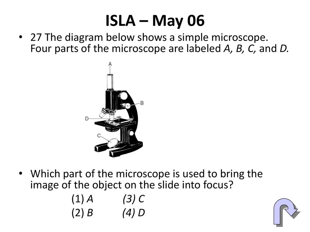
45 simple microscope diagram with labels
Light Microscope- Definition, Principle, Types, Parts, Labeled Diagram ... A light microscope is a biology laboratory instrument or tool, that uses visible light to detect and magnify very small objects and enlarge them. They use lenses to focus light on the specimen, magnifying it thus producing an image. The specimen is normally placed close to the microscopic lens. Anatomy Chart - How to Make Medical Drawings and Illustrations Pathologic anatomy focuses on how diseases affect and change the human body. Histology studies microscopic anatomy such as tissues and cells visible only under a microscope. Anatomy charts serve two main purposes: education in the form of anatomy worksheets and presentation in the form of simple healthcare illustrations.
› cells › bactcellInteractive Bacteria Cell Model - CELLS alive Ribosomes: Ribosomes give the cytoplasm of bacteria a granular appearance in electron micrographs.Though smaller than the ribosomes in eukaryotic cells, these inclusions have a similar function in translating the genetic message in messenger RNA into the production of peptide sequences (proteins).
Simple microscope diagram with labels
Free Microscope Worksheets for Simple Science Fun for Your Students 1. Parts of a Microscope . The first worksheet labels the different parts of a microscope, including the base, slide holder, and condenser. If you have a microscope, compare and contrast this worksheet to it.Also, your kids can color this microscope diagram in and read the words to each part of the microscope. Simple Microscope- Definition, Principle, Magnification, Parts ... A simple microscope is one that uses a single lens for magnification, such as a magnifying glass while a compound microscope uses several lenses to enhance the magnification of an object. It uses a lens to enlarge an object through angular magnification alone, giving the viewer an erect enlarged virtual image. The use of a single convex lens or ... alex.state.al.us › plansALEX | Alabama Learning Exchange Students will use a Venn diagram to compare lightning and static electricity. Then, students will experiment with static electricity and read nonfiction passages about lightning and lightning rods. Finally, they will apply their learning to construct a model of a lightning rod system that protects a house from a lightning-induced fire.
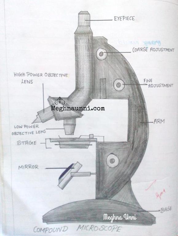
Simple microscope diagram with labels. Compound Microscope - Diagram (Parts labelled), Principle and Uses See: Labeled Diagram showing differences between compound and simple microscope parts Structural Components. The three structural components include. 1. Head. This is the upper part of the microscope that houses the optical parts. 2. Arm . This part connects the head with the base and provides stability to the microscope. Microscope Diagram - cell division of e coli with continuous media flow ... Microscope Diagram - 15 images - give a well labelled diagram of compound microscope using of typical, bio tem biological transmission electron microscope university, labelled microscope diagram gcse micropedia, a compound microscope diagram micropedia, Parts of the Microscope with Labeling (also Free Printouts) 5. Knobs (fine and coarse) By adjusting the knob, you can adjust the focus of the microscope. The majority of the microscope models today have the knobs mounted on the same part of the device. Image 5: The circled parts of the microscope are the fine and coarse adjustment knobs. Picture Source: bp.blogspot.com. › microorganisms-friend-and-foeMicroorganisms: Friend and Foe Class 8 Extra Questions ... Oct 11, 2019 · Pull out a gram or bean plant from the field. Observe its roots. You will find round struc¬tures called root nodules on the roots. Draw a diagram of the root and show the root nod¬ules. Answer: Question 2. Collect the labels from the bottles of jams and jellie on the labels. Answer: Do it yourself. Question 3. Visit a dcotor.
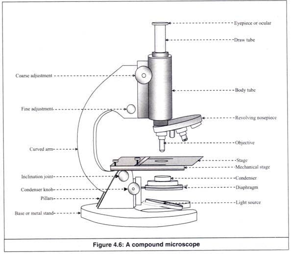
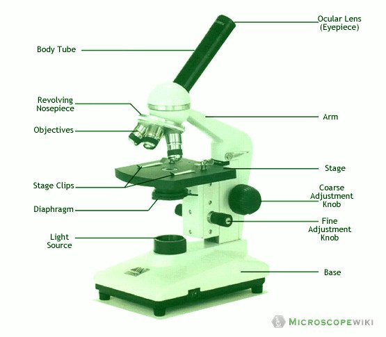
en.wikipedia.org › wiki › FluorescenceFluorescence - Wikipedia Fluorescence is the emission of light by a substance that has absorbed light or other electromagnetic radiation.It is a form of luminescence.In most cases, the emitted light has a longer wavelength, and therefore a lower photon energy, than the absorbed radiation. Microscope Parts, Function, & Labeled Diagram - slidingmotion Diaphragm. The diaphragm is also called as iris. This iris situates below the stage of the microscope. The function of the diaphragm is to control the amount of light that focuses on the specimen. This diaphragm can adjust the amount of light and intensity of light that falls on the specimen. In some standard and high-quality microscopes, this ... GCSE Science: Required practical activities - AQA Using a light microscope to observe, draw and label cells in an onion skin . Materials . In addition to access to general laboratory equipment, each student needs: • a small piece of onion • a knife or scalpel • a white tile • forceps • a microscope slide • a coverslip • a microscope • iodine solution in a dropping bottle. Compound Microscope- Definition, Labeled Diagram, Principle, Parts, Uses The optical microscope often referred to as the light microscope, is a type of microscope that uses visible light and a system of lenses to magnify images of small subjects. There are two basic types of optical microscopes: Simple microscopes. Compound microscopes. The term "compound" in compound microscopes refers to the microscope having ...
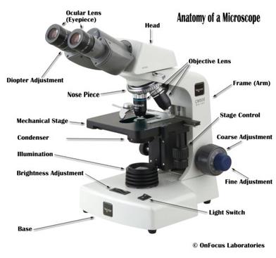
› image_maps › 32-marineMarine ecosystem — Science Learning Hub Clicking on the labels will bring up short video and images that can be used to explore marine ecosystems in greater detail. Use this interactive with the article Marine food webs . Excellent interactive for all, it would work brilliantly for all learners regardless of all reading abilities! Microscope, Microscope Parts, Labeled Diagram, and Functions Revolving Nosepiece or Turret: Turret is the part of the microscope that holds two or multiple objective lenses and helps to rotate objective lenses and also helps to easily change power. Objective Lenses: Three are 3 or 4 objective lenses on a microscope. The objective lenses almost always consist of 4x, 10x, 40x and 100x powers. The most common eyepiece lens is 10x and when it coupled with ... Interactive Bacteria Cell Model Periplasmic Space: This cellular compartment is found only in those bacteria that have both an outer membrane and plasma membrane (e.g. Gram negative bacteria).In the space are enzymes and other proteins that help digest and move nutrients into the cell. Cell Wall: Composed of peptidoglycan (polysaccharides + protein), the cell wall maintains the overall shape of a … Sperm Under Microscope with Labeled Diagram - AnatomyLearner Sperm under microscope 400x labeled. I will show you the sperm under a microscope 400x with the labeled diagram. Here in the diagram, you will see some seminiferous tubules lined by the thick germinal epithelium. The picture shows the dark Type A and pale Type B spermatogonia located at the seminiferous tubules' basal part.
Label Cell Parts | Plant & Animal Cell Activity | StoryboardThat
Marine ecosystem — Science Learning Hub Explore this interactive diagram to learn more about life in the sea. Click on the different labels to view short video clips or images about different parts of the ... It drives a carbon cycle, which involves a series of fairly simple chemical equations, and it causes the pH [level] of the sea to go down. That means the sea becomes a ...

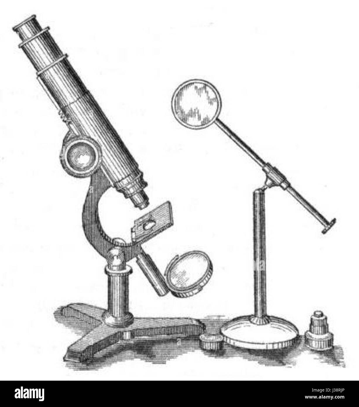
discovering the simple microscope
Hot and Cold Packs: A Thermochemistry Activity | Carolina.com Diagram your hot or cold pack. Include labels to indicate sizes and quantities of materials used. List all materials and quantities needed to create your thermal pack. Explain the steps that you will follow to build your thermal pack. Describe the safety precautions you will use when creating and testing the thermal pack.
Simple Microscope: Working Principle, Uses, Parts, and their ...
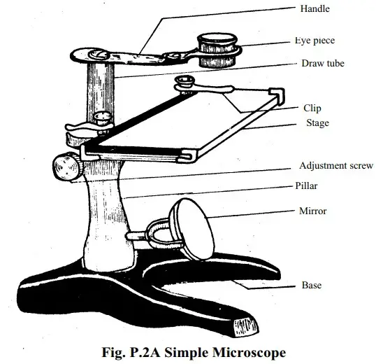
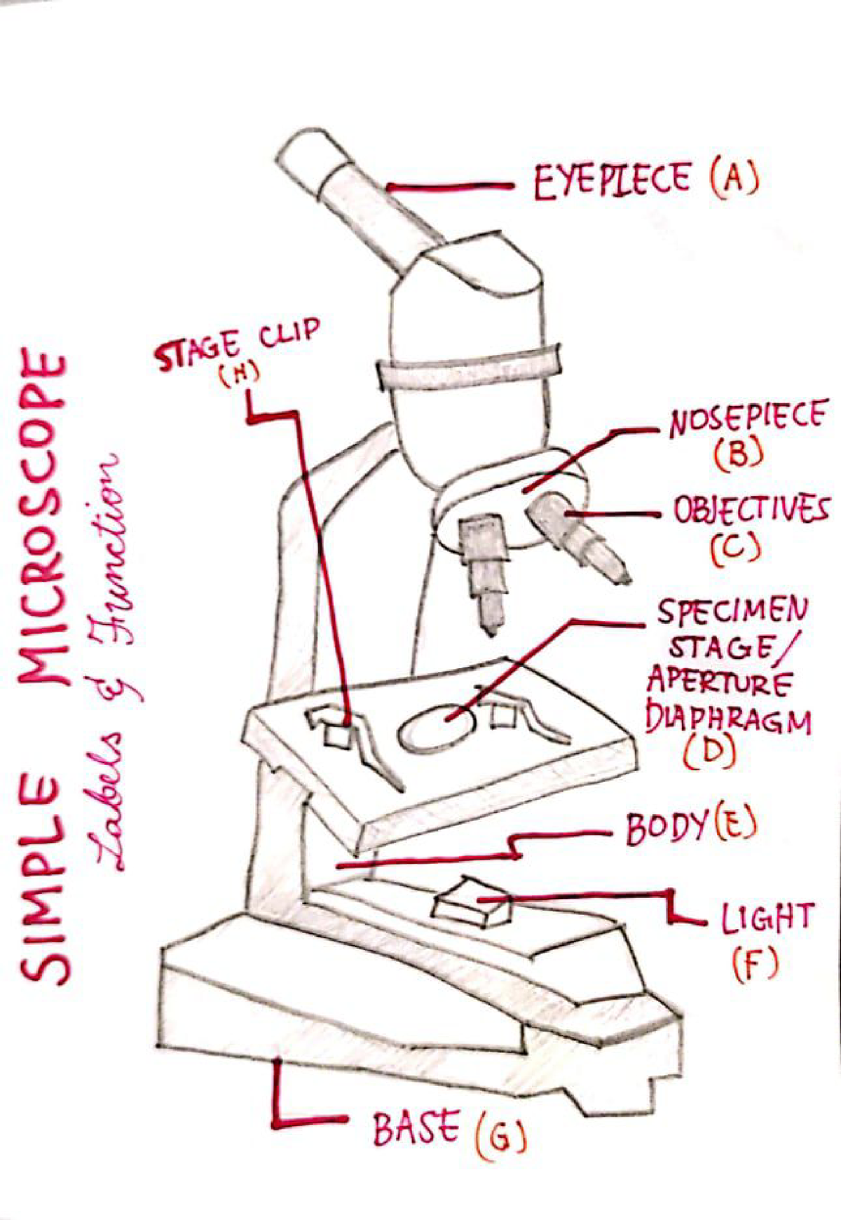
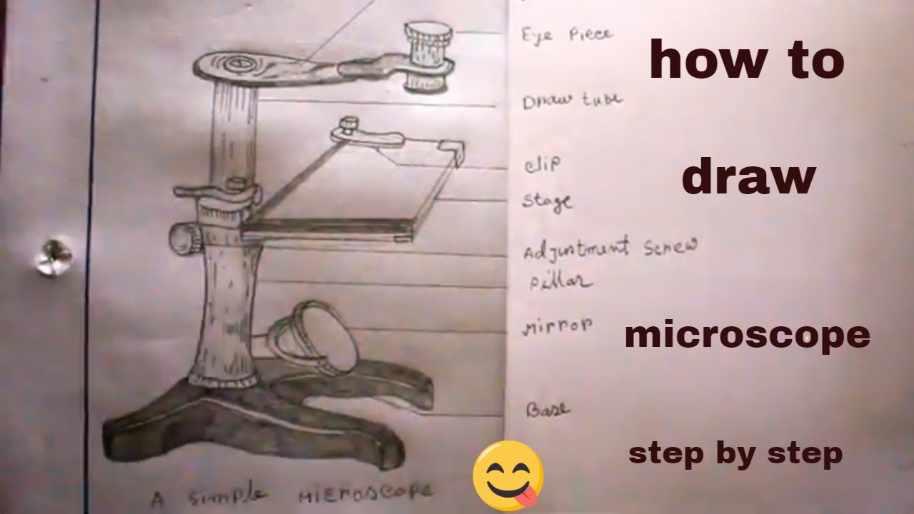
Simple Microscope - Diagram (Parts labelled), Principle, Formula and Uses Simple microscope is a magnification apparatus that uses a combination of double convex lens to form an enlarged, erect image of a specimen. The working principle of a simple microscope is that when a lens is held close to the eye, a virtual, magnified and erect image of a specimen is formed at the least possible distance from which a human eye ...

microscope | Types, Parts, History, Diagram, & Facts | Britannica
Electron Microscope Principle, Uses, Types and Images (Labeled Diagram ... Ans: A light microscope has a low resolving power (0.25µm to 0.3µm) while the electron microscope has a resolution power about 250 times higher than the light microscope at about 0.001µm. Similarly, a light microscope has a magnification of 500X to 1500x while the electron microscope has a much higher magnification of 100,000X to 300,000X.
Microscopy: Intro to microscopes & how they work (article ...
› anatomy-chartAnatomy Chart - How to Make Medical Drawings and Illustrations Pathologic anatomy focuses on how diseases affect and change the human body. Histology studies microscopic anatomy such as tissues and cells visible only under a microscope. Anatomy charts serve two main purposes: education in the form of anatomy worksheets and presentation in the form of simple healthcare illustrations.
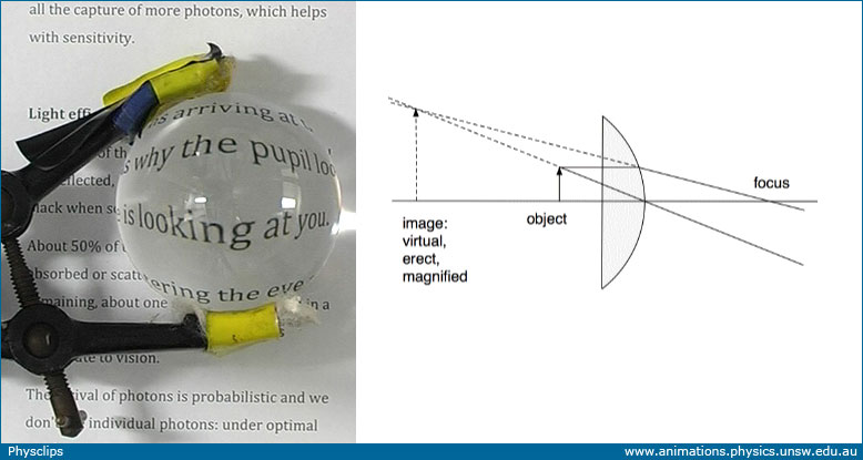
Microscopes and magnifiers: Physclips - Light
Microscope Types (with labeled diagrams) and Functions Has a higher level of magnification - Typically up to 2000x. Is used in hospitals and forensic labs by scientists, biologists and researchers to study micro organisms. Compound microscope labeled diagram. Compound microscope functions: It finds great application in areas of pathology, pedology, forensics etc.

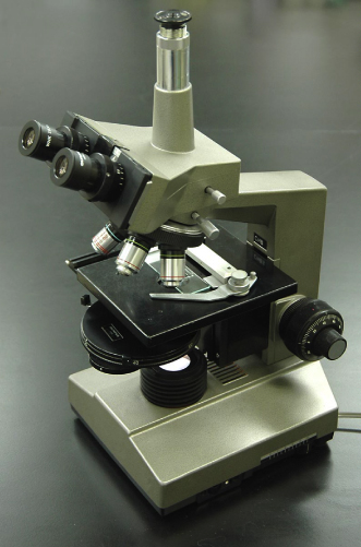
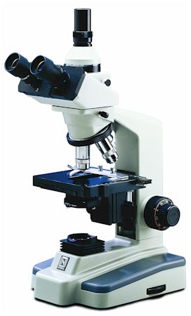
OM136C 40X-400X Student Compound Microscope
Simple Microscope Diagram, Formula, Definition, Discoverd by - adda247 Simple Microscope Diagram A Simple Microscope Simple Microscope Formula. Simple Microscope Formula is denoted as M = 1 + D/F, where D is the shortest distance of distinct vision and F is the focal length of the convex lens. Noted Fact: The higher the magnifying power of the microscope, the shorter the focal length of the lens. ...

Compound Microscope Parts – Labeled Diagram and their ...
Basic Microscope Diagram - microscope diagram purposegames, images 01 ... Basic Microscope Diagram - 15 images - label the neuron clip art at vector clip art online, microscope diagram fill online printable fillable blank pdffiller, animal anatomy biology4isc, images 01 introduction and terminology basic human anatomy,
Biology : Compound Microscope Diagram for Class 8 ...
K To 12 Science Grade 7 Learners Material - Module Read and do the activities in the section on How to Use The Light Microscope before performing Activity 2. Activity 2 Investigating plant cells Objectives In this activity, you should be able to: 1. prepare a wet mount; 2. describe a plant cell observed under the …
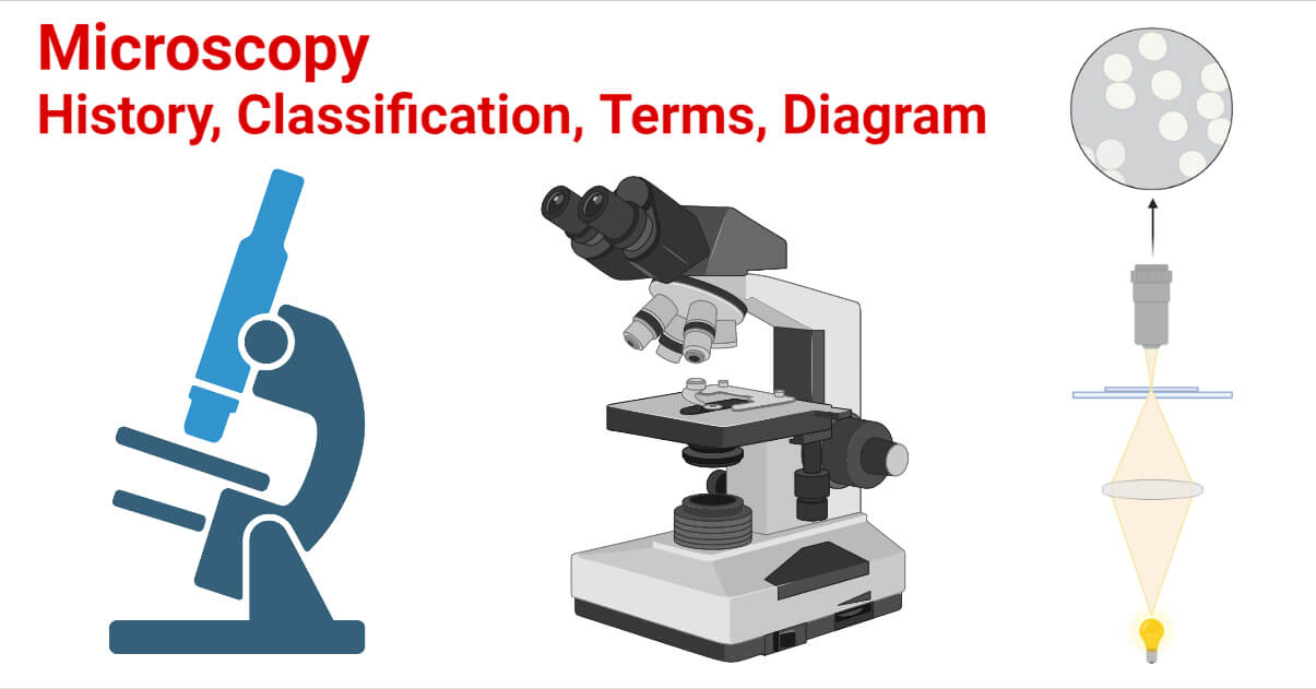
Microscopy- History, Classification, Terms, Diagram
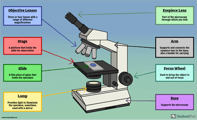
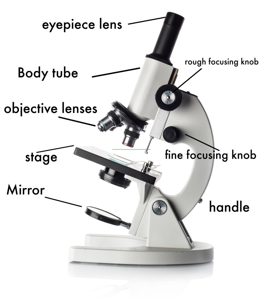
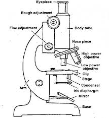
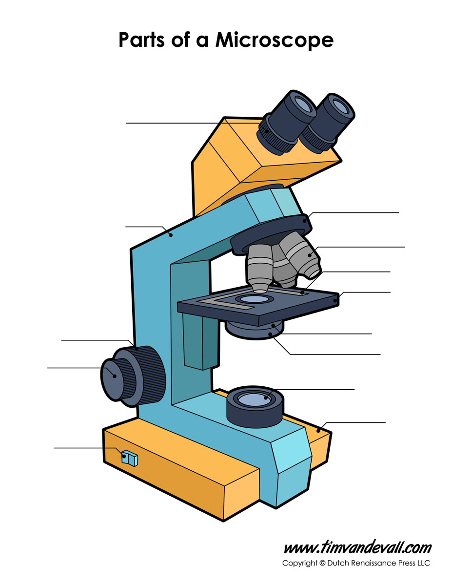
Parts of a microscope with functions and labeled diagram - Microbe Notes Head - This is also known as the body. It carries the optical parts in the upper part of the microscope. Base - It acts as microscopes support. It also carries microscopic illuminators. Arms - This is the part connecting the base and to the head and the eyepiece tube to the base of the microscope.
The Microscope Parts and Use
Simple Microscope - Parts, Functions, Diagram and Labelling Parts of the optical parts are as follows: Mirror - A simple microscope has a plano-convex mirror and its primary function is to focus the surrounding light on the object being examined. Lens - The biconvex lens is placed above the stage and its function is to magnify the size of the object being examined.

Label the microscope — Science Learning Hub
Autoclave - Wikipedia A medical autoclave is a device that uses steam to sterilize equipment and other objects. This means that all bacteria, viruses, fungi, and spores are inactivated. However, prions, such as those associated with Creutzfeldt–Jakob disease, and some toxins released by certain bacteria, such as Cereulide, may not be destroyed by autoclaving at the typical 134 °C for three minutes or 121 …
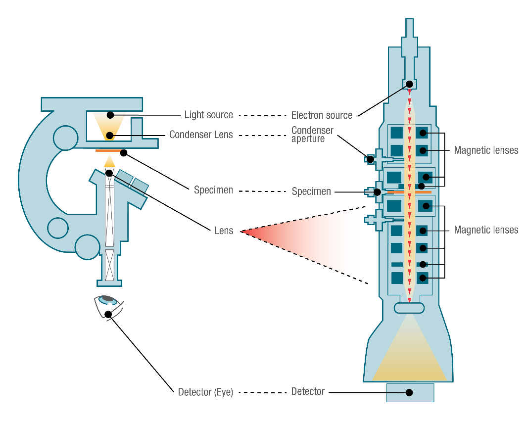
Light Microscope vs Electron Microscope - Life in Atomic ...
rockyourhomeschool.net › microscope-worksheetsFree Microscope Worksheets for Simple Science Fun for Your ... Parts of a Microscope . The first worksheet labels the different parts of a microscope, including the base, slide holder, and condenser. If you have a microscope, compare and contrast this worksheet to it. Also, your kids can color this microscope diagram in and read the words to each part of the microscope.
Microscope- Simple-AND Compound-WITH- Label - BS in Education ...
ALEX | Alabama Learning Exchange Subject: Digital Literacy and Computer Science (4), Science (4) Title: Using Code to Create an Animated Animal Description: Students will use the free online coding program, Scratch, to learn the basics of coding and how to use blocks and animations to create an animated animal. Students will show how an animated animal will receive, process, and respond to information …

Compound Microscope Parts – Labeled Diagram and their ...
Learn CBSE - Microorganisms: Friend and Foe Class 8 Extra … 11.10.2019 · Pull out a gram or bean plant from the field. Observe its roots. You will find round struc¬tures called root nodules on the roots. Draw a diagram of the root and show the root nod¬ules. Answer: Question 2. Collect the labels from the bottles of jams and jellie on the labels. Answer: Do it yourself. Question 3. Visit a dcotor.

label microscope diagram | Charts | Microscope, Anatomy bones ...
Simple Squamous Epithelium under a Microscope with a Labeled Diagram ... Simple columnar epithelium labeled. This is a labeled diagram of a simple columnar epithelium under a light microscope. I tried to show you both ciliated and nonciliated simple columnar epithelium. These diagrams show the cilia on the cell surface, rectangular cell, and elongated nucleus.
Free Microscope Drawing, Download Free Microscope Drawing png ...
Plant Cell: Diagram, Types and Functions - Embibe Exams Despite the fact that plant and animal cells are both eukaryotic and share a few cell organelles, plant cells perform different roles than animal cells. When the cells are inspected under an electron microscope, some of these changes become obvious. In this article read more about Plant Cell, Diagram, Functions, and Types.
Teaching a Lesson LE Lesson Using Regents Diagrams - ppt download
Microscope: Parts Of A Microscope With Functions And Labeled Diagram. Figure: A diagram of a microscope's components. The microscope has three basic components: the head, the base, and the arm. Head:Occasionally, the head is considered the body. It holds the optical components of the upper part of the microscope. Base:The microscope's base provides great support.
Differences between Light Microscope and Electron Microscope
Compound Microscope Diagram Labeled - parts of a microscope ... Here are a number of highest rated Compound Microscope Diagram Labeled pictures upon internet. We identified it from trustworthy source. Its submitted by supervision in the best field. We tolerate this kind of Compound Microscope Diagram Labeled graphic could possibly be the most trending topic taking into consideration we part it in google ...

The Compound Light Microscope Diagram | Quizlet
alex.state.al.us › plansALEX | Alabama Learning Exchange Students will use a Venn diagram to compare lightning and static electricity. Then, students will experiment with static electricity and read nonfiction passages about lightning and lightning rods. Finally, they will apply their learning to construct a model of a lightning rod system that protects a house from a lightning-induced fire.

easy compound microscope diagram - Clip Art Library
Simple Microscope- Definition, Principle, Magnification, Parts ... A simple microscope is one that uses a single lens for magnification, such as a magnifying glass while a compound microscope uses several lenses to enhance the magnification of an object. It uses a lens to enlarge an object through angular magnification alone, giving the viewer an erect enlarged virtual image. The use of a single convex lens or ...
Building a Fully-Functional Microscope From Lego Bricks and a ...
Free Microscope Worksheets for Simple Science Fun for Your Students 1. Parts of a Microscope . The first worksheet labels the different parts of a microscope, including the base, slide holder, and condenser. If you have a microscope, compare and contrast this worksheet to it.Also, your kids can color this microscope diagram in and read the words to each part of the microscope.
Compound Microscope. Label the numbered parts of the ...

Microscopy: History, Types of Microscope, Diagram - Embibe
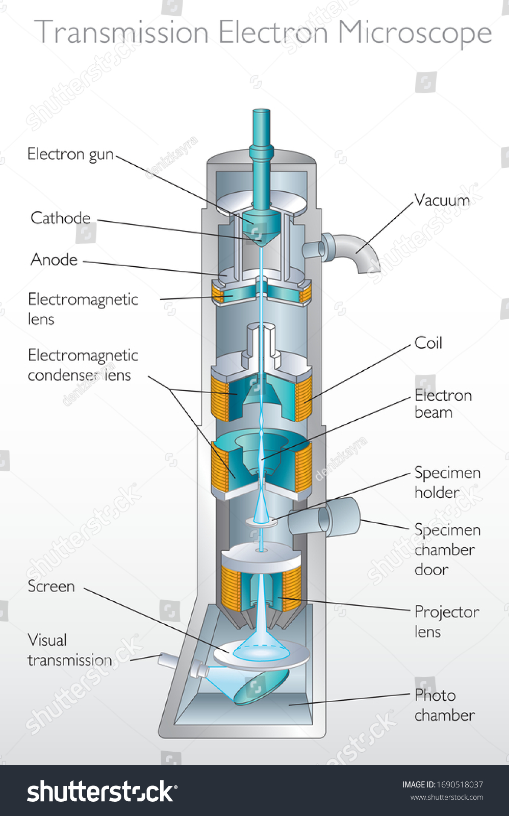
Diagram Transmission Electron Microscope Quantum Physics ...
Schematic flow diagram of a Scanning Electron Microscope ...
How to Use a Microscope
SOLVED:In what situation(s) would a transmission electron ...

Parts of a Microscope - SmartSchool Systems
PHYSICS Form 3 Topic 4 | School Base-Online
:max_bytes(150000):strip_icc()/microscopecolor3-58b978735f9b58af5c495abe.png)
Learn About Microscopes With Fun, Free Printables

Simple Microscope Definition, Magnification, Parts And Uses
How TO Draw simple microscope step by step/simple microscope drawing/for science project

Draw a neat labeled diagram for the formation of an class 12 ...
Compound and Stereo- microscopes - Microscopes 4 Schools

Simple Microscope - Parts, Functions, Diagram and Labelling ...

Label the microscope — Science Learning Hub
Microscope Diagram Labeled, Unlabeled and Blank | Parts of a ...
microscope - Kids | Britannica Kids | Homework Help
Microscope Types (with labeled diagrams) and Functions
Optical microscope - Wikipedia

Compound Microscope Parts, Functions, and Labeled Diagram ...
Microscope Parts and Functions
Compound microscope Black and White Stock Photos & Images - Alamy

Compound Microscope Parts, Functions, and Labeled Diagram ...
Post a Comment for "45 simple microscope diagram with labels"